Special Hours this Week:
TUESDAY 2/24 – 9:00am to 6:00pm
WEDNESDAY 2/25 – CLOSED
THURSDAY 2/26 – 8:30am to 6:00pm
FRIDAY 2/27 – 8:30am to 5:00pm

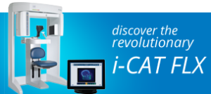
Our commitment to patient’s treatment and safety has kept us at the cutting edge of best technology available for orthodontic diagnosis. Adhering to the ALARA principle (As Low As Reasonably Achievable) to reduce radiation exposure, Bridgham Barr Orthodontics has invested in the newest radiographic technology and are currently the only orthodontic practice in Westchester and Putnam counties to have the i-CAT FLX.
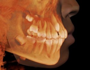
This state of the art cone beam technology enables us to capture more detailed three-dimensional information than traditional two-dimensional orthodontic radiographs (panoramic and cephalometric “x-rays”), at less than half the radiation exposure*. One 3D orthodontic scan will collect the information necessary to produce all the diagnostic images (panoramic, cephalometric, TMJ panel) at 11 uSv, which is less radiation than most digital panoramic x-ray machines. Therefore, our patients can expect 1/8 to 1/2 the amount of radiation that a typical orthodontic patient will undergo at another office using standard digital technology (and our image provides far more diagnostic information)! The images precisely capture accurate views of jaws, teeth, roots, TMJ, airway and sinuses without magnification or distortion. This technology allows us to evaluate jaw size as well as precisely locate impacted and unerupted teeth with adjustable cross-sectional views and volume renderings.
 Drs. Bridgham and Barr understand that a better diagnosis means better treatment for our patients. More accurate and highly defined information about the teeth, jaws, and facial complex allows us to develop the most efficient and effective treatment plan for each patient, which is then carried through to successful case completion.
Patient safety and comfort is a top priority at Bridgham Barr Orthodontics. This advanced 3D technology allows the doctors to easily select the appropriate scan for each patient at the lowest acceptable radiation dose. The award winning i-CAT FLX has an i-Collimator that electronically adjusts the field-of-view to limit radiation only to the area of scanning interest, so that our clear 3D images can be customized for each patient, depending on the amount of information required at a specific stage of treatment. We believe that the ability to have maximum control over the dose of radiation delivered to our patients is of critical importance to people of all ages, as well as clinically responsible care.
*Ludlow JB, Walker C. Assessment of phantom dosimetry and image quality of i-CAT FLX cone-beam computed tomography. Am J Orthod Dentofacial Orthop. 2013;144(6):802-817.

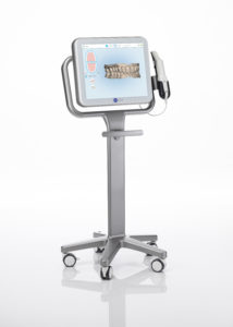
We are proud to offer our patients the iTero® Element digital impression system, by Align Technology. Our digital impression system replaces the uncomfortable, unpleasant-tasting, messy and sometimes inaccurate traditional putty impressions. No bulky trays or sticky putty needed!
We use the Element digital scanners to capture images of your tooth surfaces and gum tissue. You can follow the scanning progress on the screen to see your 3D model being formed. These impressions will help the doctors formulate a personalized treatment plan that meets your specific orthodontic needs. We can also use them to visually show you your treatment options and help us keep you up-to-date on your treatment progress. Not only is a digital impression more comfortable, it’s also much more accurate. The scanner takes an incredibly detailed impression of your teeth and gums and eliminates the need for retaking impressions.
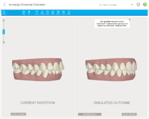
 We can even use the Invisalign Outcome Simulator to visualize how your teeth may look after treatment. Seeing the final result before you decide can take the guess work out of your decision and start you on your journey to the smile you’ve always wanted!